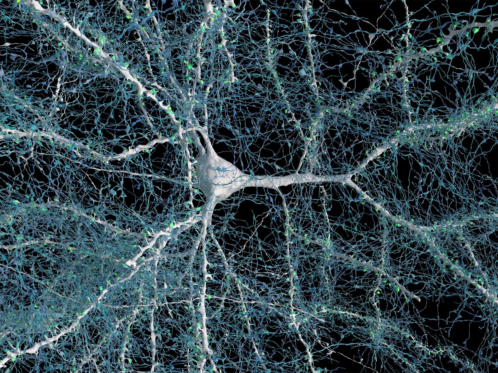
A single neuron is shown with 5,600 of the nerve fibers (blue) that connect to it. The synapses that make these connections are in green. Credit: Google Research & Lichtman Lab, Harvard University. Renderings by D. Berger, Harvard
Researchers used high-resolution electron microscopy to image a small piece of human brain tissue, generating a 3D map of over 57,000 cells and nearly 150 million synapses. Their findings reveal intricate details about cell types and connections, highlighting the complexity of the brain and advancing the field of connectomics.
- Researchers generated a high-resolution map of all the cells and connections in a single cubic millimeter of the human brain.
- The results reveal previously unseen details of brain structure and provide a resource for further studies.
Fully understanding how the human brain works requires knowing the relationships between the various cells that make up the brain. This entails visualizing the brain’s structure on the scale of nanometers in order to see the connections between neurons.
Imaging Techniques and Research Methodology
A team of researchers, led by Dr. Jeff Lichtman at Harvard University and Dr. Viren Jain at Google Research, used electron microscopy (EM) to image a cubic millimeter-sized piece of human brain tissue at high resolution. The tissue was removed from the cerebral cortex of a patient as part of a surgery for epilepsy.
The team began by cutting the tissue into more than 5,000 slices, or sections, each of which was then imaged by EM. This yielded about 1.4 petabytes, or 1,400 terabytes, of data. Using these data, the researchers generated a 3D reconstruction of almost every cell in the sample. Results of the NIH-funded study were published in the journal Science.

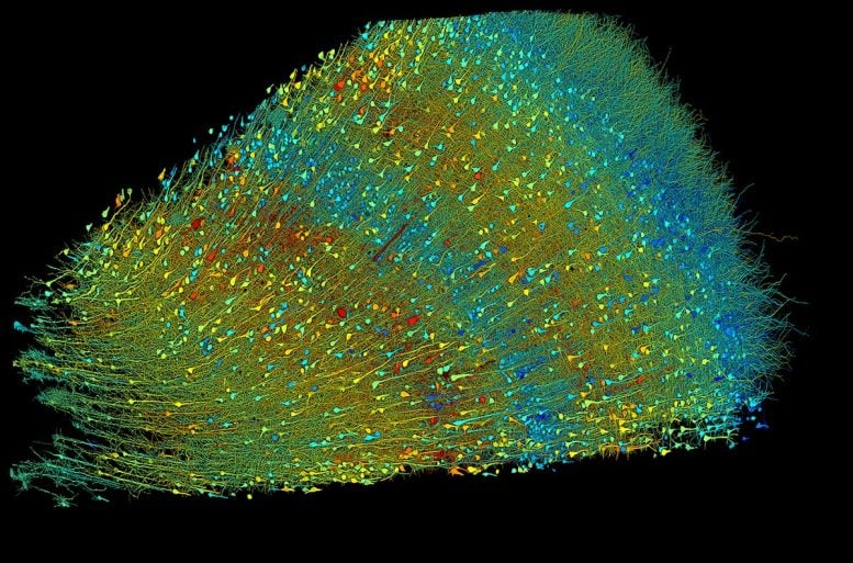
Researchers built a 3D image of nearly every neuron and its connections within a small piece of human brain tissue. This image shows six layers of neurons, colorized according to the size of each cell’s central core. Credit: Google Research & Lichtman Lab, Harvard University. Renderings by D. Berger, Harvard
Detailed Findings and Cellular Analysis
Analysis of individual cells in the sample revealed a total of more than 57,000 cells. Most of these were either neurons, which send electrical signals, or glia, which provide various support functions to the neurons. Glia outnumbered neurons 2-to-1. The most common glial cells were oligodendrocytes, which provide structural support and electrical insulation to neurons. The one cubic mm sample also contained about 230 mm of blood vessels.
The reconstruction revealed structural details not seen before. The researchers analyzed a type of neuron, called triangular cells, that are found in the deepest layer of the cerebral cortex. Many of these adopted one of two orientations, which were mirror images of each other. The significance of this organization remains unknown.
Synapses and Connections
The team used DOI: 10.1126/science.adk4858


Discussion about this post