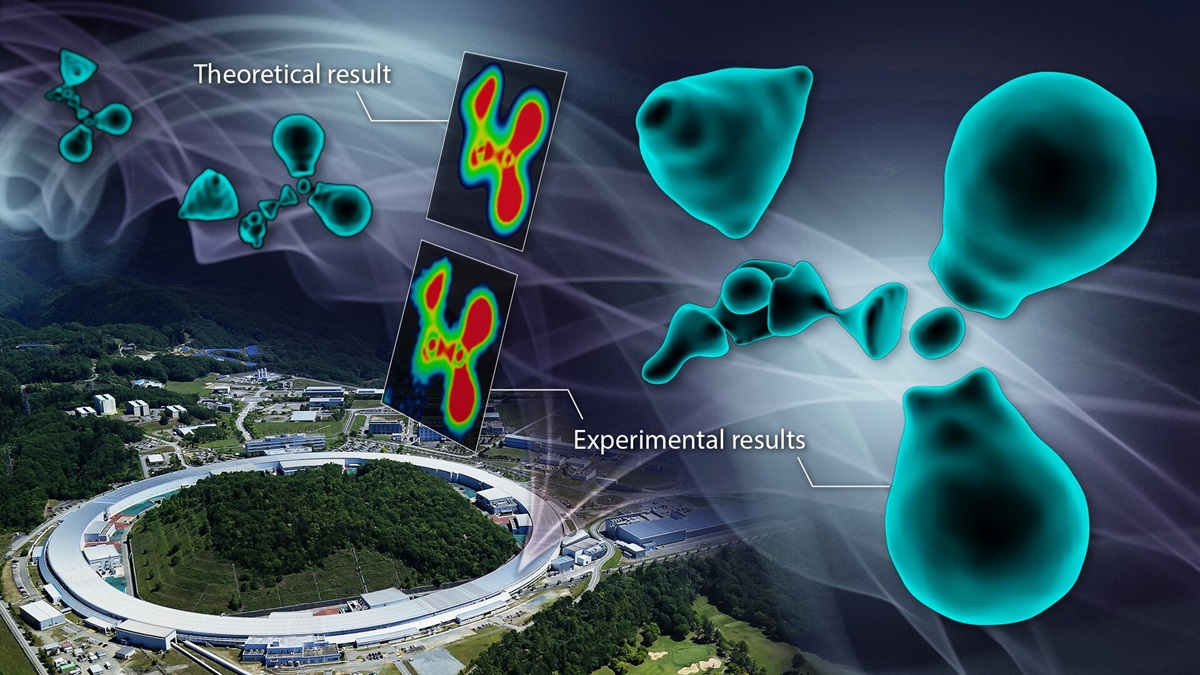
For the first time, scientists have imaged the distribution of the electrons in the outermost shell, known as valence electrons, of atoms in an organic molecule.
The experimental breakthrough sheds light on the fundamental nature of chemical bonds. It could have implications for pharmaceuticals and chemical engineering.
Behaviour of electrons in atoms depends on the orbitals they occupy in the atomic shell. These orbitals can loosely be thought of as how close the electron is orbiting the atomic nucleus.
Different conventions label these shells differently (for example, starting from the inner-most shell: 1,2,3, …; s, p, d, …; or K, L, M, …).
Electrons closer to the nucleus, called inner-shell or core electrons, provide stabilisation and don’t interact with other atoms.
Outer shell electrons, however, describe the material’s properties including how the atoms chemically bond with other atoms. The number of valence electrons depends on the element’s position in the periodic table and specific rules about the number of electrons which can fit into each orbital shell.
As you go through the periodic table you see an increase in the number of electrons in an atom. For example, carbon is the 6th element in the periodic table and, when not ionically charged, has 6 electrons. Two of these go into the first shell and the other 4 go into the second shell which can hold a maximum of 8 electrons before the third shell starts getting filled. Their 4 valence electrons make carbon atoms very receptive to chemical bonding.
Getting information about valence electrons, though, is a challenge. Until now, researchers have had to rely on theoretical models and spectroscopy to estimate valence electron behaviour.
In the new study, published in the Journal of the American Chemical Society, the researchers used synchrotron X-ray diffraction experiments to image only the valence electrons.
The experiments were done at the Super Photon ring-8 GeV facility in Japan.
“Using this method, we observed the electron state of the glycine molecule, a type of amino acid,” says corresponding author Hiroshi Sawa from Nagoya University. “Although the method was relatively simple to perform, the result was impressive.”
Sawa says the valence electrons appeared as separate blobs, contrary to expectations.
“The observed electron cloud did not exhibit the smooth, enveloping shape that many predicted, but rather a fragmented, discrete state.”
Translating the image to a colour map, the researchers found interruptions in the electron distribution around carbon atoms in the molecule.
“When carbon forms bonds with surrounding atoms, it reconstructs its electron cloud to create hybridised orbitals,” Sawa explains.
Because of this hybridisation and the wave-like nature of electrons, instead of a continuous ‘cloud’ of electrons, there are parts of the hybrid orbitals where no electrons exist.
This surprising result was confirmed through quantum chemical calculations done by collaborators at Hokkaido University.
“I believe it has been helpful in providing a clear conclusion to the ambiguous understanding of bonding states that has puzzled researchers since the 19th century,” Sawa says. “Visualising electron behaviour is a challenging endeavour.”
“This study has made it possible to directly visualise the essence of chemical bonds, potentially contributing to the design of functional materials and the understanding of reaction mechanisms,” Sawa adds. “This is because it aids in discussing the electronic states of molecules, which are difficult to infer from just the chemical structural formula.
“It may, for example, explain why some medicines work and others don’t. Fields where interactions influence functionality and structural stability, such as organic semiconductors and research on the structure of DNA double helixes, are likely to benefit most from our research.
“I believe our findings have astonished many researchers and validated the model proposed by quantum chemistry.”


Discussion about this post