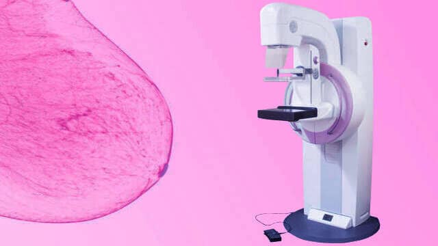
Breast cancer is second common cause of death among the women’s between 40 to 55 and here mammography machine comes in picture.
It is advisable to women’s to perform mammography screening after 40 year age on the regular basis because there are high chances of breast cancer during this period.
Due to family history, body changes, reproductive or menstrual history, obesity, breast density, after menopause less physical activity, unbalance diet, previous biopsy are the few common reasons.
Screening on regular basis helps to detect any kind of malignancy in it’s initial stage to start treatment thus improve fair chances to cure breast cancer.
What is Mammography?
It is a process to achieve good quality inside images (mammogram) of breasts using x-ray to increase the radiologist’s ability to observe abnormal masses.
Although in some cases instead of x-ray ultrasound used for breast study but most common practice is mammography machine.
It expose the breasts to a relatively small amount of radiation, typically less than 20% of average yearly
background radiation. Mammography is of two type, screening and diagnostic.
A screening mammography is the standard screening test for breast cancer now days. A “Breast screening” exam is a test used for routine check-ups, to make sure that presumably healthy women do not have a specific disease.
Once a mass is found on a screening mammogram, the patient will often return to have a diagnostic
mammogram, which consists of specialized, close-up views of the mass with extra compression.
This will help the radiologist better characterize the mass as either benign or malignant.
Mammography Machine Technology
Equipment evolved over at least the last 40 years to the present. While there are some differences from one manufacturer to another, there are also many characteristics and features that are common to all.

Basically it is a C-arm with fixes source to detector distance with some modifications and extra movements. That is what we will introduce and cover here.
X-ray Tube Anode
Whereas most x-ray tubes use tungsten as the anode material, mammography equipment uses molybdenum anodes or in some designs, a dual material anode with an additional rhodium track.
These materials are used because they produce a characteristic radiation spectrum that is close to optimum for breast imaging.
Filter
X-ray machines use aluminum or “aluminum equivalent” to filter the x-ray beam (soft x-ray) to reduce unnecessary exposure to the patient.
Mammography uses filters that work on a different principle and are used to enhance contrast sensitivity. Molybdenum (same as in the anode) is the standard filter material.
Some systems allow the operator (or automatic control function) to select either the molybdenum or a rhodium filter to optimize the spectrum for specific breast conditions.
Focal Spots
The typical x-ray tube for mammography has two selectable focal spots. The spots are generally smaller than for other x-ray procedures.
Because of the requirements for minimal blurring and good visibility of detail to see the small calcifications. The smaller of the two spots is generally used for the magnification technique.
Compression Device
Good compression of the breast is one of the essentials of effective mammography (and a common source of patient discomfort and concern). Potential benefits derived from compression include:
A more uniform breast thickness resulting in a better fit of the exposure into the film latitude or dynamic range.
1 Reduced blurring from patient motion.
2 Reduced scattered radiation and improved contrast sensitivity.
3 Reduced radiation dose.
4 Better visualization of tissues near the chest wall.
Grid
A grid is used as in other x-ray procedures to absorb scattered radiation and improve contrast sensitivity.
Compared to grids for general x-ray imaging, grids for mammography have a lower ratio and the material between the strips is selected for low x-ray absorption.
The grid is contained in a Bucky device that moves it during the x-ray exposure to blur and reduce the visibility of the grid lines.
Detector / Sensor / Receptor
Both film/screen and digital detectors are used for mammography. Each has special characteristics to enhance image quality.
To complete study about X-Ray fundamentals and it’s application follow X-Ray.
Digital Mammography
Digital mammography has several advantages over film for optimizing the contrast transfer from the breast to the image display and the maximizing the overall contrast sensitivity.
The major advantages are here.
Digital Receptor Dynamic Range
A valuable characteristic of most digital receptors is a constant sensitivity over a wide range of exposures. This is very different from the relatively narrow latitude or dynamic range of film.
The full exposure histogram will be easily covered by the wide dynamic range and that is a considerable variation in exposure to the receptor (exposure error) can be tolerated without loss of contrast.
The transfer of exposure contrast into digital image contrast is represented by a linear (straight-line) rather than the steep characteristic curve of film with its limited latitude.
The digital image recorded by the typical digital receptor will have relatively low contrast, but it will be uniform over the full exposure range.
The next step is to select the exposure range representing the actual image, that is the histogram, and to enhance the contrast by digital processing and windowing.
Digital Image Processing
One of the great advantages of digital imaging is the ability to apply a variety of processing procedures to change the image characteristics to improve quality and visibility.
Contrast processing is common in most forms of digital radiography and is used to make the digitally acquired radiographs like more conventional film radiographs with respect to contrast.
The advantage is that the user can select from many different “film characteristics” to meet the needs of specific clinical procedures.
For example, in general radiography, one “characteristic curve” type would be appropriate for chest imaging while another would be used for imaging the extremities.
In digital mammography the various contrast processing procedures are generally built into the system and might vary to some extent from one manufacturer to another.
Windowing
Windowing used in the display and viewing of most digital images (including CT, MRI, etc) is the last step in optimizing the contrast and visibility of specific objects and structures within an image.
The various contrast characteristics of digital imaging (wide dynamic range, processing, and windowing) can be combined to produce maximum contrast sensitivity as required in mammography.
3D Mammography/DBT
Reviewing breast tissue slice by slice allows the radiologist to view breast tissue in a way never before possible.

Digital Breast Tomosynthesis (DBT) is a 3D imaging technology that acquires a series of low-dose projection images of the compressed breast at different angles.
The system produces thin slices of the breast, allowing radiologists to step through slices one-millimeter at a time.
Unlike a traditional mammogram, the slices separate objects at different heights in the breast, producing a 3-dimensional image.
X-ray tube moves in an arc across the breast. A series of low dose images are acquired from different angles. Total dose approximately the same as one 2D digital mammogram.
Projection images are reconstructed into 1 mm slices utilizes low-level x-rays to produce multiple images of the breast, layer by layer, using a swinging camera.
This layering of images makes it simpler to detect normal breast structures (milk ducts, lobules, fatty tissues, etc.) from cancerous ones.
X-rays are converted into limited 3-D digital images for radiologists to examine. Computer Aided Detection (CAD) assists in spotting regions where cancer seems to be present.
Dense tissue is more easily examined through Tomography than traditional Mammography increases detection of Invasive breast cancers by 40% in comparison to 2D mammography.
To know about CT how it works or reconstruct body internal organ images using x-ray follow ct scan.
For diy projects follow and subscribe our Computernxtechnology youtube channel.


Discussion about this post